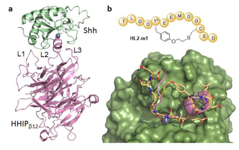
Figure 2.

Macrocyclic HHIP L2 loop mimic. (a) Crystal structure of Shh (green) in complex with the extracellular domain of HHIP (pink) (pdb 3HO525a). The three loop regions of HHIP involved in Shh binding are labeled and the zinc ion in the L2 binding cleft of Shh is shown as sphere model (blue). (b) Top: schematic structure of the macrocyclic peptide HL2-m1. Bottom: model of HL2-m1 (yellow, stick model) bound to Shh (green, surface model). The L2 loop of HHIP (pink, ribbon model) is superimposed to the modeled complex.
