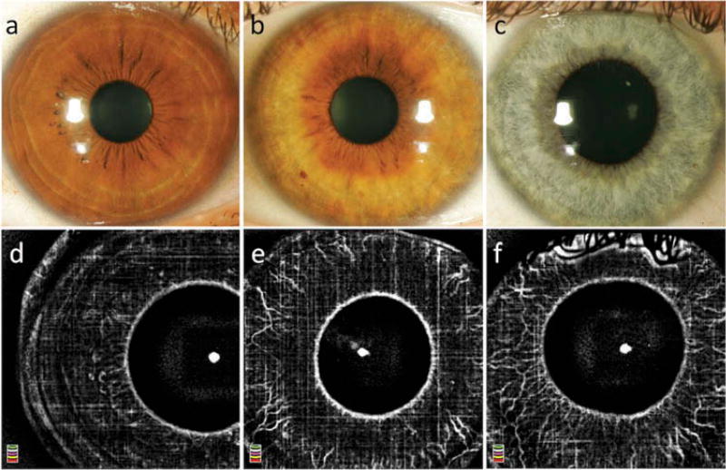
Figure 1.
Slit lamp photography (a–c) and optical coherence tomography angiography (d–f) of eyes without NVI with dark (a,d), medium (b,e), and light (c,f) iris pigmentation. Note the influence of pigment density on the detection of the physiological iris vasculature. In the iris with dense pigmentation (= brown iris; a,d) few blood vessels are visualized. In the medium pigmented iris (= hazel; b,e), the vessels are observed; however, a larger number of vessels and more details of the healthy vasculature are visualized in lightly pigmented iris (= blue; c,f).

