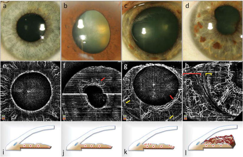
Figure 3.
Slit lamp photographs (a–d) and optical coherence tomography angiographic (OCTA) imaging of neovascularization of the iris (NVI) (e–h) and corresponding schematic cross-sections (i–l) based on the clinical stages described by Gartner and Henkind.2 Healthy iris vasculature (a, e) with physiological radial vessel configuration of large-caliber stromal vessels (i). Stage 1 NVI: a ring of fine blood vessels around the pupillary border, which was clinically invisible (b), can be observed in en face OCTA (red arrow in f). The corresponding schematic (j) illustrates the independent growth of fine new blood vessels at the pupillary border and at the iris root. Vessel growth at the iris root was not observed in this eye. Stage 2 NVI: blood vessels around the pupil (red arrow in g) and at the root of the iris (yellow arrows in g) are thicker and more tortuous (c, k); however, there are hardly any connections between the two neovascular networks. Image (c) illustrates the clinical appearance. Stage 3 NVI: connections between the peripupillary NVI network and the iris root network have formed (d, h, l). The vessels are very tortuous and show the same thickness as physiological iris vasculature. A network of neovascularization of the iris root can be observed (yellow bracket in h). Conjunctival vessels are highlighted by a red bracket in (h). The bright white horizontal line in (f) is caused by motion artifact.

