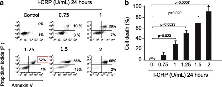
Fig. 2.

Phosphatidylserine exposure and membrane permeability of HeLa cells after I-CRP exposure. a Cell death was measured by flow cytometry through Annexin-V and PI staining in HeLa cells treated with different concentrations (0.75, 1.0, 1.25 1.5, 2 U/mL) of I-CRP for 24 h. The percentages refer to Annexin-V-positive/PI-negative or Annexin-V-positive/PI-positive staining analyzed by flowjo software. b Cells were treated and analyzed as in (A) and graphed
