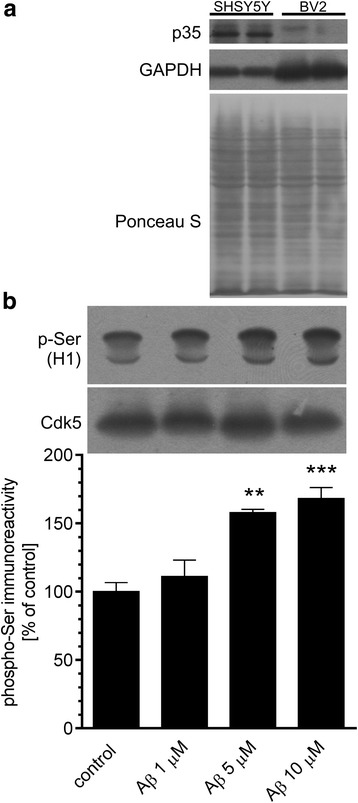
Fig. 8.

Aβ administration evokes Cdk5 activation in neuronal cells. a Immunoreactivity of p35 and GAPDH in SH-SY5Y and BV2 cells was analysed 3 h after Aβ treatment by SDS-PAGE and Western blotting. Representative pictures were shown. PonceauS staining was used as a loading control. b In SH-SY5Y cells treated with 1, 5 or 10 μM Aβ for 3 h, Cdk5 kinase activity was measured as described under “Experimental Procedures.” Results of densitometric analysis of phosphorylated histone H1 are presented as the mean ± SEM from four independent experiments (n = 4). **, ***p < 0.01 and 0.001 compared to control, using a one-way ANOVA followed by the Bonferroni test
