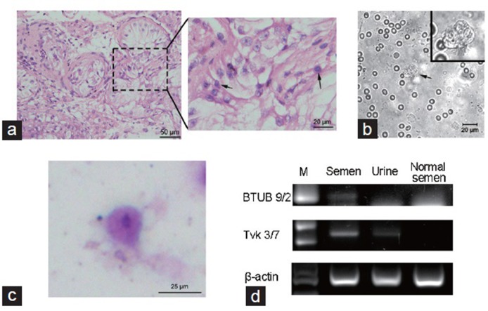
Figure 1.
(a) HE staining of testicular biopsies, showed a severe disruption in spermatogenesis. Arrows indicate the germ cells. Scale bars = 50 μm (left) and 20 μm (right). (b) Wet preparation of testicular biopsies, showed a Trichomonas-like flagellate (arrow). Scale bar = 20 μm. (c) Wright-Giemsa staining of the wet preparation smear, arrow indicates Trichomonas vaginalis. Scale bar = 25 μm. (d) PCR analysis of Trichomonas vaginalis from the semen and urine. A normozoospermic semen (normal semen) sample was applied as negative control. M: 100 bp DNA ladder. HE: Hematoxylin-Eosin; PCR: polymerase chain reaction.

