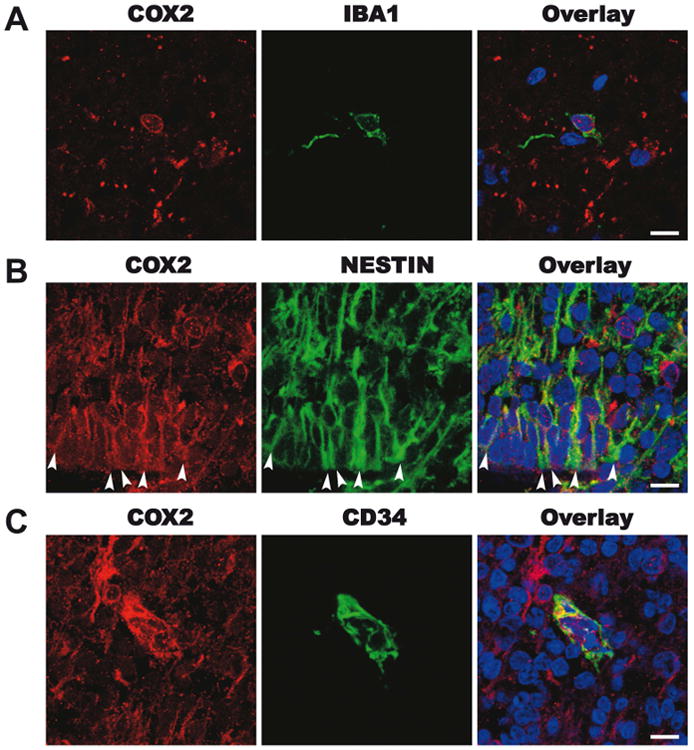
Figure 1. COX2 immunohistochemistry in the human third trimester brain.

Representative images from the dorsal cortex of a 30-week human fetal brain. A. In the subplate, red COX2, green IBA1+ microglia and an overlay panel including DAPI positive nuclear staining. B. in the subventricular zone, red COX2, green nestin+ putative radial glia and astrocytes and an overlay panel including DAPI+ nuclear staining. C. In the subventricular zone, red COX2, green CD34+ endothelia cell and an overlay panel including DAPI positive nuclear staining. Scale bar = 10μm.
