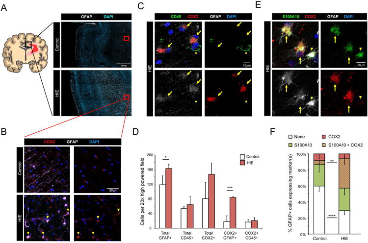
Figure 2. COX2 immunohistochemistry of subcortical white matter from human hypoxic ischemic encephalopathy (HIE) cases.

A Cartoon illustrating affected white matter areas in human term HIE. Black box represents cingulate region used for analysis. Red boxes are examples of subcortical white matter regions used for analysis. HIE cases exhibit increased GFAP (white) immunoreactivity. B. Representative images from term infants with or without HIE, stained for COX2 (red) and GFAP (white). Arrowheads mark COX2+ GFAP+ astrocytes. C. Representative images of white matter expression of COX2 in GFAP+ astrocytes. Arrows mark COX2+ GFAP+ astrocytes. Arrowhead marks a COX2- CD45+ microglia/myeloid cell. D. Quantification of indicated cell types in control and HIE white matter. E & F. Representative images (E) and quantification (F) of S100A10 co-expression with COX2 in white matter GFAP+ astrocytes. Arrows mark GFAP+ astrocytes co-expressing S100A10 and COX2. Arrowhead marks a GFAP+ astrocytes expressing only S100A10. Data from n=4 control and n=3 HIE cases. p-values calculated from two-tailed unpaired t-tested. * p <0.05, ** p<0.01, *** p<0.005, **** p<0.001
