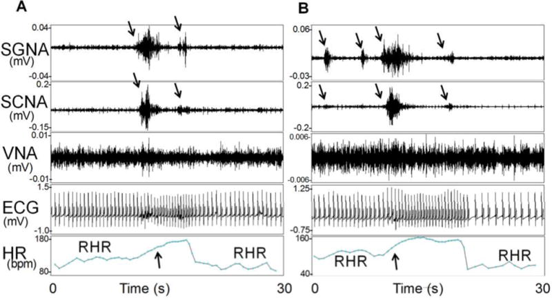
Figure 1.

SCNA and SGNA are associated with heart rate elevation in an ambulatory dog. The first portion of Panel A shows rhythmic heart rate (HR) variations consistent with respiratory heart rate responses (RHR). SGNA and SCNA (downward arrows) then activated simultaneously, resulting in heart rate acceleration (upward arrow). There were no obvious changes of VNA in this recording. Simultaneous cessation of the SGNA and SCNA was associated with a reduction of the heart rate and the resumption of RHR. B shows simultaneous activation of SGNA, SCNA (downward arrows) in the same dog 25 seconds after Panel A. Downward arrows point to simultaneous nerve activities in SGNA and SCNA. Upward arrow indicates the onset of tachycardia. ECG, electrocardiogram. (From Robinson et al, J Cardiovasc Electrophysiol; 2015)(33)
