
Figure 3.
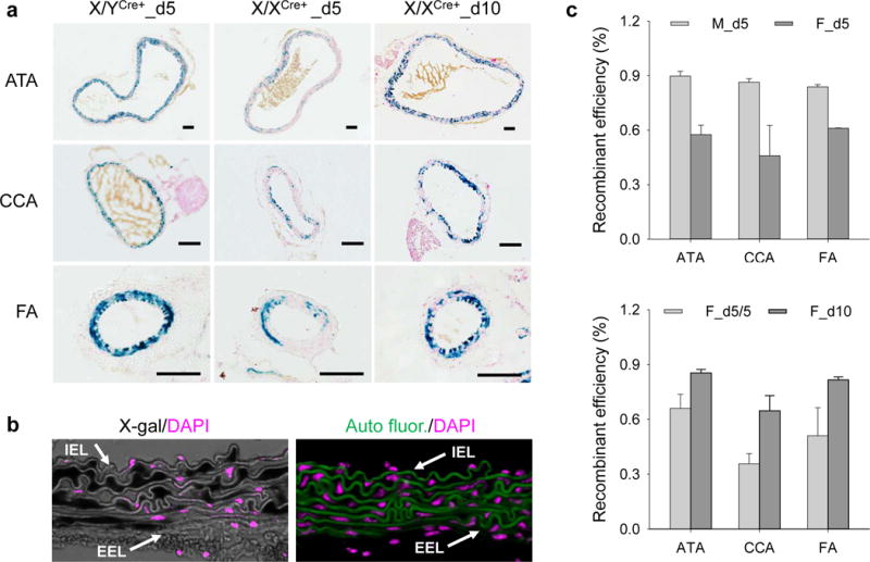
The X/XCre+ mouse line is less efficient than the X/YCre+ mouse line, but its efficiency can be enhanced by prolonged tamoxifen induction. (a) X-gal staining of arteries with indicated anatomic location, genotype, and duration of tamoxifen induction. Blue: positive X-gal staining; red: nuclear fast red counterstain; scale bars: 50 μm. (b) An example of imaging techniques utilized for assessing the recombinant efficiency. Left panel: X-gal staining (dark black) with DAPI counterstain (pink) of an ATA sample. Note the resistance of X-gal positive cells to DAPI counterstain. Right panel: DAPI counterstain of the section adjacent to that assigned to X-gal staining. Green: autofluorescence; pink: DAPI counterstain. Arrows point to internal and external elastic laminae (IEL and EEL, respectively). (c) Recombinant efficiency in muscular arteries. M and F represent X/YCre+ and X/XCre+ mice, respectively. Symbols d5, d10, and d5/5 indicate respectively 5, 10, and 5 tamoxifen doses for a duration of 5, 10, and 5 plus 5 days respectively. Data were analyzed using two-way ANOVA. M_d5 vs. F_d5: P<0.001; F_d5/5 vs. F_d10: P<0.001.