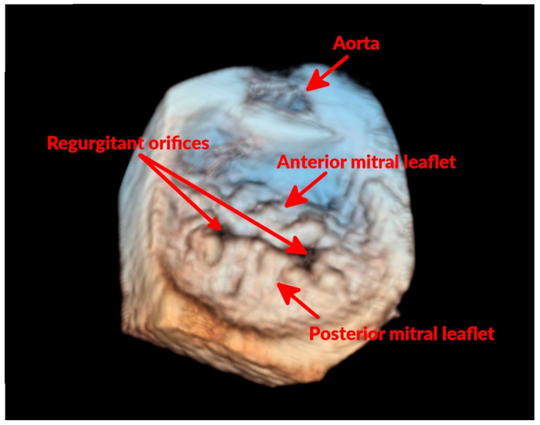
Figure 3.
Three-dimensional echocardiographic “surgical” view of the MV of a dog with mitral prolapse. In this three-dimensional echocardiographic image, the MV is visualized as seen from the LA. It can be noticed how several areas of both anterior and posterior MV leaflets are bulging. It can also be noticed that the two leaflets fail to coapt in two areas (regurgitant orifices), which is where the MR occurs.

