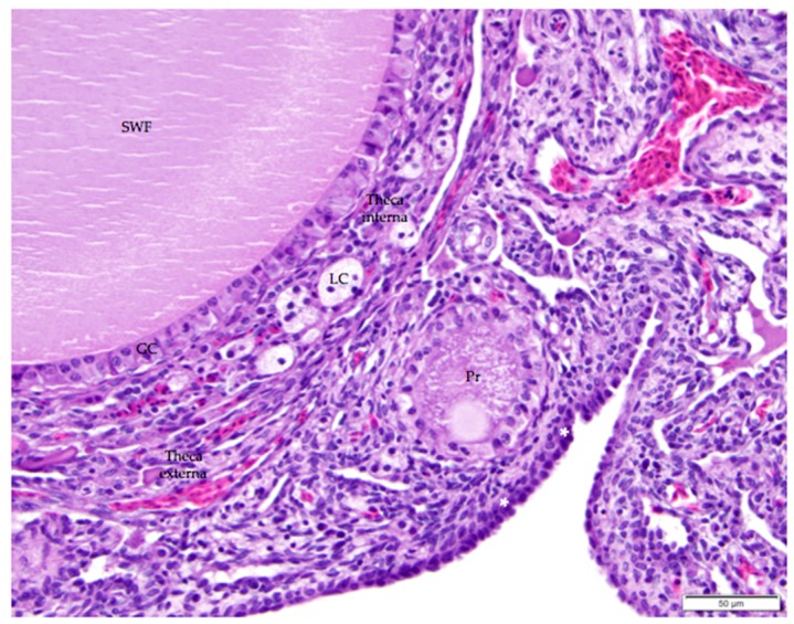
Figure 7.
The thin, collagenous tunica albuginea that separates the surface epithelium from the ovarian cortex is occasionally visible (white asterisks). The image includes a small white follicle (SWF) and a primordial follicle (Pr). A single layer of granulosa cells (GC) delineates each follicle. The small white follicle is also surrounded by theca interna and theca externa layers. Luteal cell clusters (LC) are visible in the theca interna layer. H & E. 400×.

