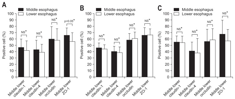
Fig. 2.
A comparison of the percentages of cells that stained positively for each tight junction protein in the middle and lower esophagus. (A) Patients with proton pump inhibitor refractory gastroesophageal reflux disease symptoms (n=62). There were no significant differences in the proportions of cells positive for claudin-1, claudin-4, or occludin between the middle and lower esophagus. The percentage of zonula occludin-1 (ZO-1)-positive cells, however, was significantly lower in the lower than in the middle esophagus (56.0%±14.0% vs 66.0%±11.5%, p<0.05). (B) Healthy controls (n=10). There were no significant differences between the proportion of cells positive for claudin-1, claudin-4, occludin, or ZO-1 in the middle and lower esophagus. (C) Patients with eosinophilic esophagitis (EoE) (n=6). There were no significant differences in the proportion of cells positive for claudin-1, claudin-4, occludin, or ZO-1 between the middle and lower esophagus.
NS, not significant. *Unpaired t-test.

