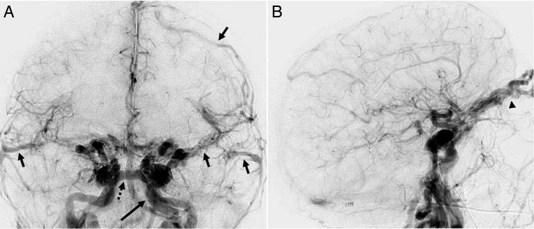
Figure 3.
Biplane intravenous digital subtraction angiography in the frontal (A) and lateral projections (B) in the early arterial phase demonstrating marked arteriovenous shunting and early venous drainage to the left cavernous sinus via the intercavernous sinus (dotted arrow) and cortical venous reflux (arrows). A left petrous internal carotid artery pseudoaneurysm is incidentally noted (long arrow). Additional early venous drainage and venous hypertension is noted within the bilaterally dilated superior ophthalmic veins (arrowhead).

