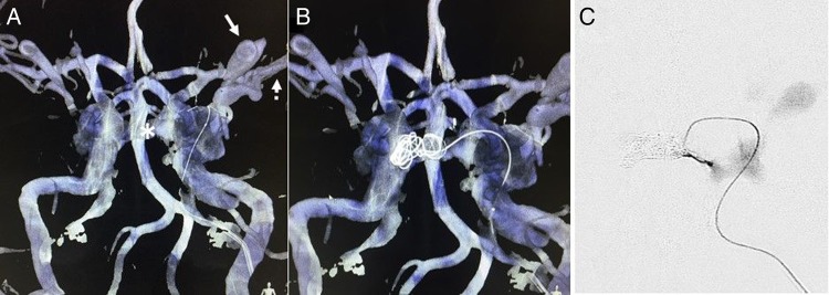
Figure 4.
The TAILOREd technique demonstrating overlay images of live fluoroscopy on the preoperative MR angiography (in blue) and cone beam CT (in white). (A) The microcatheter is navigated through the ipsilateral cavernous sinus into the contralateral cavernous sinus via the intercavernous sinus (asterisk). The ipsilateral superior ophthalmic vein can also clearly be seen on the three-dimensional roadmap (arrow) as well as the cortical venous reflux (dotted arrow). (B) Coils are placed within the contralateral cavernous sinus and intercavernous sinus. (C) Microcatheter injection confirms occlusion of the intercavernous sinus.

