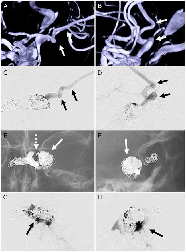
Figure 5.
Anteroposterior (A and C) and lateral (B and D) three-dimensional roadmap and microcatheter runs, respectively, demonstrating catheterization of a venous pouch with direct posterior lateral cortical venous reflux through the sphenoparietal sinus and sylvian vein (arrows). This high-risk feature was embolized with coils (arrows), with additional coils deployed within the cavernous sinus adjacent to the fistula as shown on anteroposterior (E) and lateral fluoroscopy (F). The left internal carotid artery cavernous segment is noted as a gap within the coil mass (E, dotted arrow). Anteroposterior (G) and lateral (H) final microcatheter runs of the left cavernous sinus in the late phase of the injection demonstrate stasis of contrast in the cavernous sinus proper without ophthalmic or cortical vein opacification, suggesting occlusion of the carotid–cavernous fistula.

