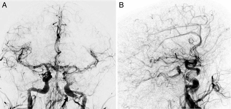
Figure 6.
Anteroposterior (A) and lateral (B) final post-embolization intravenous digital subtraction angiography confirms complete occlusion of the left direct carotid–cavernous fistula without evidence of early venous drainage. An unrelated chronic dissection and pseudoaneurysm of the left internal carotid artery is again seen (black arrow).

