
Figure 7.
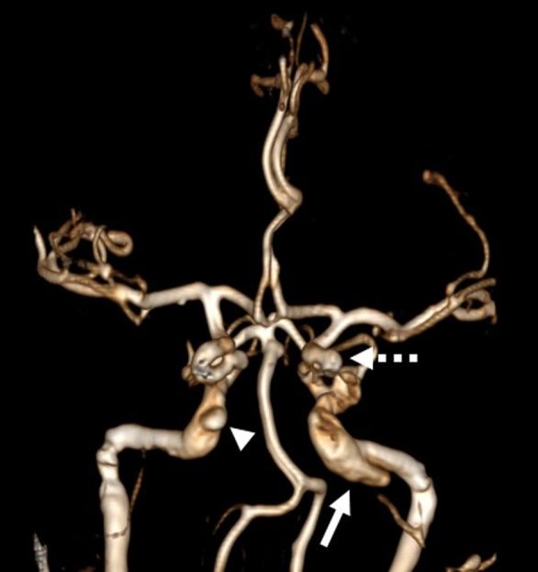
Six-week follow-up contrast-enhanced MR angiography confirms persistent occlusion of the left direct carotid–cavernous fistula with residual left cavernous (dotted arrow) and petrous (arrow) segment internal carotid artery (ICA) pseudoaneurysms. A new contralateral pseudoaneurysm is noted at the distal petrous segment of the right ICA (arrowhead).