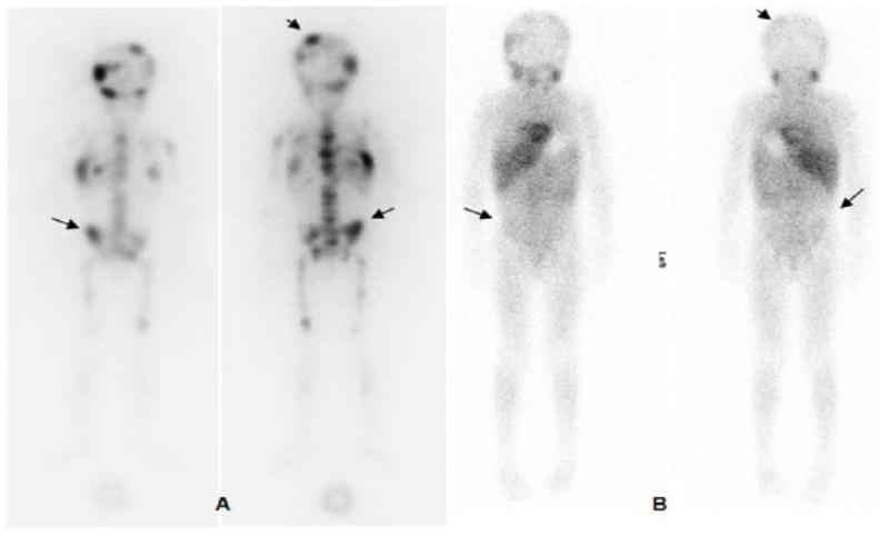
Figure 4.

123I-MIBG pre-therapy (A) and post-therapy (B) scans: anterior and posterior whole-body images performed at 24 h post-injection. Images show multiple abnormal foci of uptake in the axial and appendicular skeleton that are resolved (arrows) or decreased (arrow heads) in the post-therapy scan.
