Figure 4.
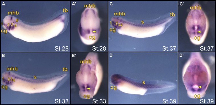
In situ hybridization of ism1 expression in late stage (St.) X. laevis embryos. Embryos are shown in a lateral (A–D) or anterior (A’–D’) view with the cement gland (cg) labeled for ventral orientation. (A) St.28 embryo showing strong expression in the branchial arches (ba), midbrain–hindbrain boundary (mhb), ear placode (e), and tailbud (tb), with the yellow arrowheads marking the primitive mouth. (B) St.33 embryos showing decreased expression in the ba, mhb, and tb, with concentrated expression surrounding the primitive mouth (arrowhead) and expression in the somites (s). (C and D) St.37 and 39 embryos showing continued concentrated ism1 expression surrounding the primitive mouth (arrowhead).

