Figure 5.

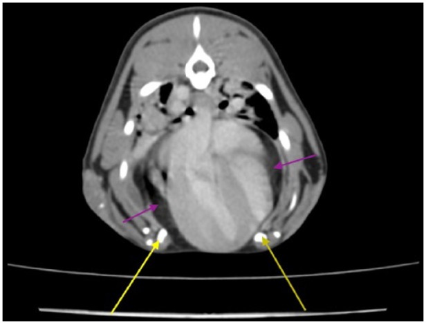
CT scan, sagittal view. The two halves of the bifid sternum are seen (yellow arrows). The peritoneopericardial hernia is seen on this view with a fat content around the heart in a distended pericardium (purple arrows)

CT scan, sagittal view. The two halves of the bifid sternum are seen (yellow arrows). The peritoneopericardial hernia is seen on this view with a fat content around the heart in a distended pericardium (purple arrows)