Fig. 5.
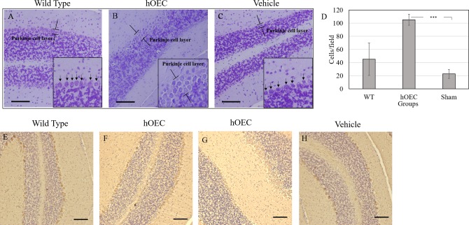
Recognizing Purkinje cells in the cerebellum. The Purkinje cells in the cerebellum were stained by crystal violet in (A) wild-type, (B) human olfactory ensheathing cell (hOEC)-transplanted, and (C) vehicle-treated groups. The number of Purkinje cells were quantified (D). The Purkinje cells in the cerebellum were recognized by their marker calbindin using immunohistochemistry (IHC) staining in (E) wild-type, (F and G) hOEC-transplanted, and (H) vehicle-treated groups. Scale bar: 100 µm. Arrows indicate the Purkinje cells.

