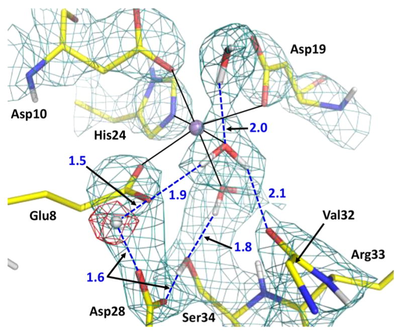
Figure 4.

Mn2+ (violet sphere)-binding site and protonation of the Asp28–Glu8 pair. The 2FO – FC neutron scattering length density map (teal mesh) is shown at the 1.5σ contour level. The omit difference FO –FC neutron scattering length density map (red mesh) for the D atom in the low-barrier hydrogen bond is shown at the 3σ contour level. Hydrogen bonds are shown as blue dashed lines, and D⋯O distances are given in units of angstroms. The side chain of Val32 was omitted for the sake of clarity.
