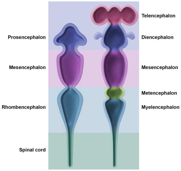
Figure 2. Main subdivisions of the embryonic mouse central nervous system.
The early embryonic brain (left image) is composed of three swellings at the cephalic end of the neural tube that develop into three primary brain vesicles: prosencephalon (forebrain), mesencephalon (midbrain), and rhombencephalon (hindbrain). Soon after (right image), the three vesicles shift their contours to assume a five-vesicle conformation. The prosencephalon divides into the telencephalon (paired cerebral hemispheres) and the diencephalon (thalamus and hypothalamus). The mesencephalon does not subdivide. The rhombencephalon partitions into the metencephalon (cerebellum and pons) and the myelencephalon (medulla oblongata). The caudal end of the neural tube develops into the spinal cord.

