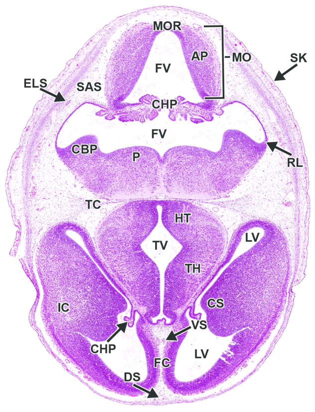
Figure 20. Representative image of the embryonic mouse brain at E14.5.
H&E-stained, transverse section. AP= alar plate; CBP= cerebellar primordium; CHP= choroid plexus; CS= corpus striatum; ELS= endolymphatic sac; FC= falx cerebri; FV= fourth ventricle; HT= hypothalamus; IC= internal capsule; LV= lateral ventricle; MO= medulla oblongata; MOR= medulla oblongata roof; P= pons; RL= rhombic lip; SAS= subarachnoid space (future); TC= tentorium cerebelli; TH= thalamus; TV= third ventricle.

