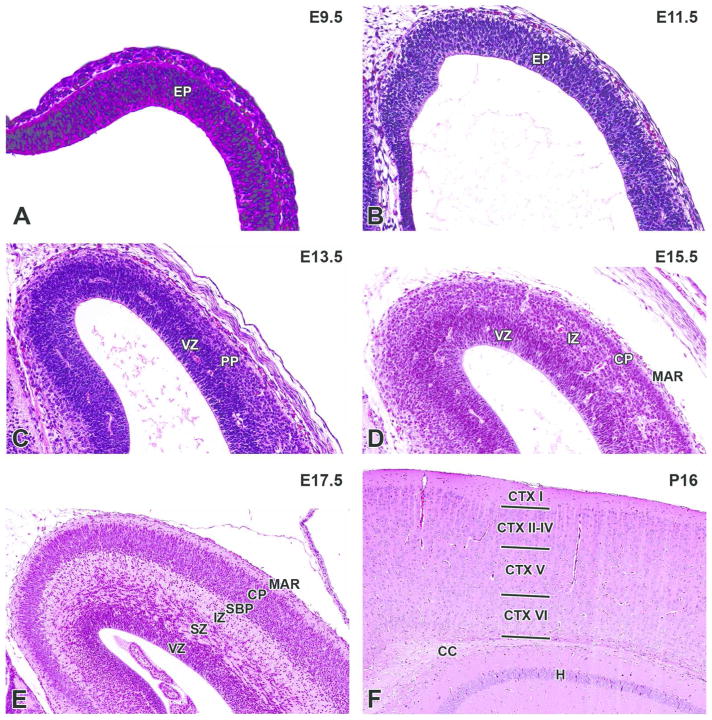
Figure 7. Representative images of the cerebral cortex during brain development.
H&E-stained sections of the prosencephalic (A) and telencephalic (B, C, D, E, F) walls. A. E9.5, sagittal section. B. E11.5, transverse section. C. E13.5, coronal section. D. E15.5, coronal section. E. E17.5, coronal section. F. P21, coronal section. CC= corpus callosum; CP= cortical plate; CTX I–VI= cortical layer I–VI; EP= ependymal layer; H= hippocampus; IZ= intermediate zone; MAR= marginal layer; PP= cortical preplate; SBP= cortical subplate; SZ= subventricular zone; VZ= ventricular zone.

