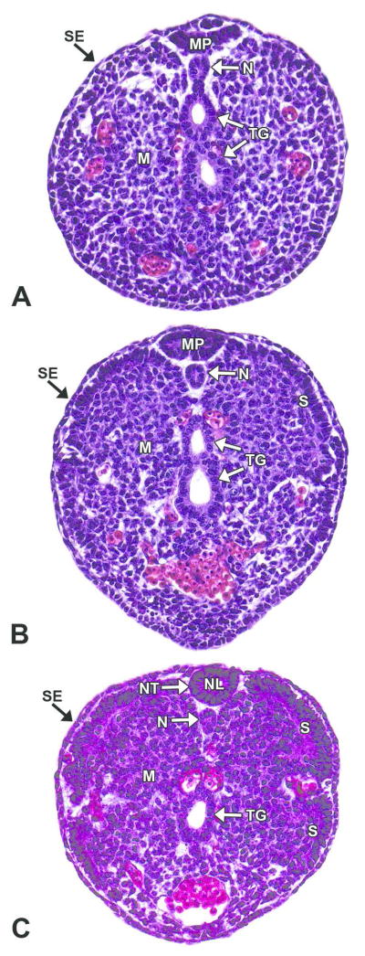
Figure 9. Representative images of secondary neurulation.
H&E-stained, transverse sections of an E12.5 embryo. Secondary neurulation forms the neural tube (NT) caudal to the caudal neuropore through the condensation of tail bud mesenchyme (M), which undergoes mesenchymal-to-epithelial transition to form a medullary plate (MP) of neuroepithelium. A, B. Caudal tail bud and mid-tail bud, respectively. The medullary plate consists of columnar epithelium from mesenchymal cells that extend dorsoventrally from the surface ectoderm (SE). C. Cranial tail bud. A slit-like lumen appears below the medullary plate, and the plate begins to round while cavitating to form the neural lumen (NL). The center then connects to the lumen of the remainder of the neural tube. The notochord (N) forms from mesenchymal cells directly ventral to the secondary neural tube. Other abbreviations: S= somite; TG= tail gut.

