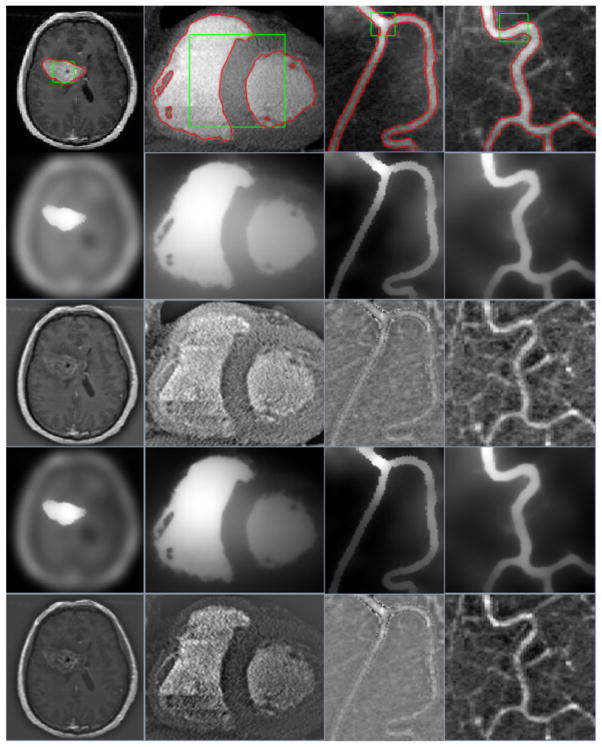
Fig. 2.
The segmentation results of our proposed model for four medical images by replacing several default parameter values from left to right (i.e., λ2 = 0.07, σ = 5, Δt = 0.01 and ε = 1.0 for the first image; σ = 19, Δt = 0.01 and ε = 7 for the second image; σ = 5, Δt = 0.35 and ε = 1 for the third image; and σ = 8, Δt = 0.01 and ε = 1 for the last image). Row 1: initial rectangle contours and their final evolutions. Row 2 and Row 3: fitted images ILFI and DLFI. Row 4 and Row 5: fitted images ISFI and DSFI.

