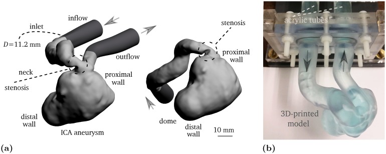
Fig 1.
(a) Lumen of the giant pre-treatment internal carotid artery aneurysm in 2:1 scale. The light gray geometry indicates the STereoLithography file taken from [24], and the dark gray regions are the extensions that connect the lumen to the acrylic tubes. (b) 3D-printed phantom in 2:1 scale filled with water-glycerol mixture infused with CuSO4. The arrows represent the flow direction.

