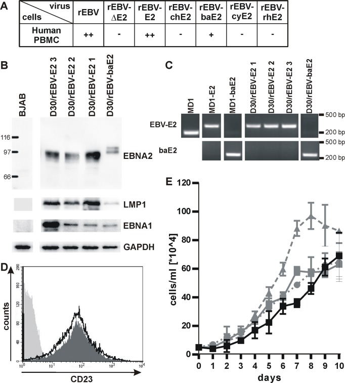
Fig 3. De novo immortalization of human PBMC by rEBVs.
A) Human PBMC were infected with rEBVs, cultured in 50 microtiter wells, and outgrowth of immortalized cells was quantified 6 weeks after infection. Average results for at least 2 experiments are shown using independent virus preparations of rEBV (n = 3), rEBVΔE2 (n = 3), rEBV-E2 (n = 5), rEBV-chE2 (n = 4), rEBV-baE2 (n = 3), rEBV-cyE2 (n = 2), and rEBV-rhE2 (n = 4). +: 1–20 wells with growth/50 total wells, ++: 21-44/50, -: no growth. B) Representative cell lines derived from PBMC of the same donor (D30) infected with rEBV-E2 and rEBV-baE2 were immunoblotted for detection of EBNA2, EBV LMP1, and EBV EBNA1. Detection of GAPDH controlled for loading and BJAB cells served as EBV-negative control. C) Expression of only one EBNA2 species was confirmed by species-specific PCR amplification for EBV or baboon LCV EBNA2 in rEBV immortalized cells. Wild type MD1, MD1-E2, and MD1-baE2 BAC DNAs purified from bacteria served as controls. D) Surface expression of the EBNA2-regulated marker CD23 on rEBV immortalized cells D30/rEBV-baE2 (dark grey fill), and D30/rEBV-E2 1 (black line) or the EBV negative cell line BJAB (light grey fill) was determined by flow cytometry. E) Growth curve of D30 LCLs transformed with rEBV-E2 (grey curves, line: clone 1, dashes: 2, dots: 3) or rEBV-baE2 (black line).

