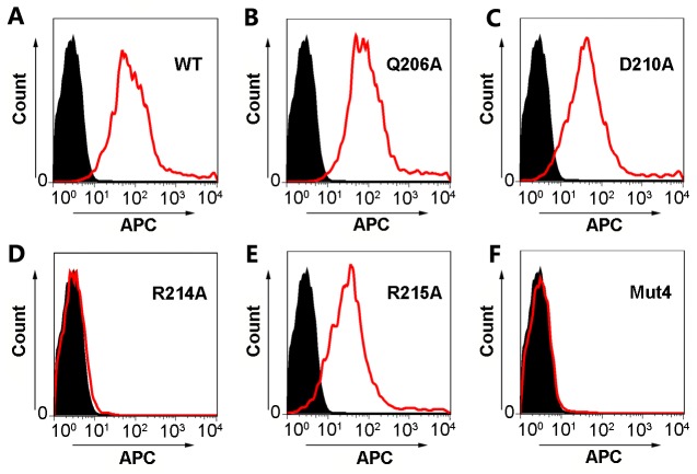
Fig 5. Flow cytometry analysis to verify the 1H1 epitope by mutagenesis.
The 293T cells were transfected by wild type (A) PRV gB or mutants (B-F) expression vectors to allow protein expression on the cell surface. The 1H1 mAb was then used to stain the transfected cells, which were further stained by APC-linked secondary antibody. The fluorescence signals of the cells were visualized and quantified by flow cytometry. The profiles of cells transfected with pEGFP-N1 empty vectors are represented by solid black areas (negative control) and the cells transfected with gB or mutant expression plasmids are represented by red silhouettes. WT, wild type; Mut4, quadruple mutants.

