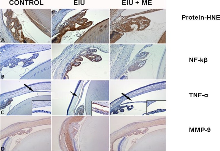
Figure 5.
Topical application of ME ameliorated several molecular markers of inflammation in the rat eye. Immunohistochemistry of the oxidative and inflammatory markers was performed as described in the text. (A) Protein-HNE adduct formation after 24 hours of LPS challenge was prominent in epithelial cells of the ciliary body and epithelial cells of the lens (×400 magnification). (B) Immunostaining of NF-kβ after 6 hours of LPS challenge was elevated in the epithelial cells of the ciliary body (×200 magnification). Secretion of TNF-α (C) was observed in the anterior chamber (×100 magnification, inset ×400) and MMP-9 (D) was observed within the cornea and sclera (×100 magnification) after 24 hours of LPS challenge. Treatment with ME decreased the expression of all of these toxic markers.

