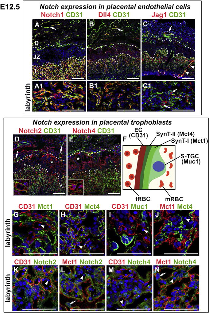
Fig. 4. Notch receptors and ligands in the E12.5 placenta.
IF was performed on tissue sections and nuclei were stained with DAPI (blue). (A–E) CD31 stains ECs of maternal vessels in the decidua and ECs of fetal vessels in the labyrinth. (A, A1) Notch1 was expressed in ECs of maternal vessels (arrow) and fetal labyrinthine vessels (A1). (B, B1) Dll4 was expressed in CD31+ ECs of maternal vessels (arrow) and fetal labyrinthine vessels. (C, C1) Jag1 (red) was expressed in CD31+ ECs of maternal vessels (arrow); in ECs (yellow signal) and perivascular cells of maternal canals (arrowheads); and in labyrinthine perivascular cells (arrows in C1). (D, E) Notch2 (D) and Notch4 (E) were expressed in decidual cells; P-TGCs (insets); and in SpTCs and GlyTCs surrounding maternal blood spaces (asterisks). Notch2 was expressed in TBs of chimeric maternal vessels (remodeled spiral arteries) at the decidua/junctional interface (arrows in D). (F) Schematic of the labyrinthine maternal-fetal interface with the markers used to identify cell types. (G–J) CD31 stains ECs, Mct1 identifies synctiotrophoblast layer SynT-I, Mct4 identifies synctiotrophoblast layer SynT-II, and Muc1 identifies S-TGCs in the labyrinth. Notch2 (K, L) and Notch4 (M, N) were not co-expressed with CD31 or Mct1. Notch2 and Notch4 were expressed in SynT-II, the synctiotrophoblast layer directly adjacent to fetal vessels, which contain nucleated red blood cells (arrowheads). Notch2 (L) and Notch4 (N) were expressed in S-TGCs (arrows). Dotted lines separate the layers of the placenta. fRBC, fetal red blood cell; mRBC, maternal red blood cell, D, decidua; JZ, junctional zone; L, labyrinth. All scale bars = 100 µm.

