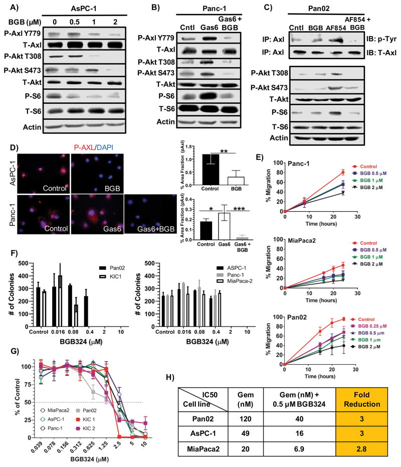
Figure 1. BGB324 inhibits Axl activity in pancreatic cancer cells in vitro.
A) AsPC-1 cells were serum starved in 1% FBS in media overnight and subsequently treated with increasing concentrations of BGB324 (BGB) for 30 minutes. Cell lysates were probed by Western blotting for the indicated targets. B) Panc-1 cells were serum starved overnight as above and subsequently stimulated with DMSO (0.02%) in media (Cntl), Gas6 (200 ng/ml) or Gas6 + BGB324 (2 μM) for 30 minutes followed by Western blot analysis for the indicated targets. C) Pan02 cells were serum starved overnight and subsequently treated with DMSO (0.02%) in media, BGB324 alone (2 μM), AF854 (2.5 nM), an Axl antibody that can stimulate Axl activation (20), or AF854 + BGB324 for 30 minutes. Axl was immunoprecipitated followed by probing for phospho-tyrosine (p-Tyr) and total Axl (upper two rows). Total cell lysates were also probed for the indicated targets. D) Phosphorylated Axl was detected in AsPC-1 and Panc-1 by immunocytochemistry. AsPC-1 cells were serum starved overnight as above (Control) then treated with BGB324 (2 μM) for 30 minutes. Panc-1 cells were serum starved and treated with Gas6 (200 ng/ml) or Gas6 + BGB324 (2 μM). AsPC-1 and Panc-1 cells were fixed permeabilized, stained with anti-phospho-Axl, and subsequently developed by immunofluorescence. Images were analyzed using Elements software; quantification of % area fraction is shown. Data are displayed as mean ± SD and represent 5 images per cell line. *P < 0.05; **P < 0.01; ***P < 0.005. E) The effect of BGB324 on cell migration was assessed by a “scratch” assay. Monolayers of the indicated cells were wounded with a pipet tip. The cells were incubated in serum-free media +/− BGB324 at the indicated concentrations. Wound closure was monitored at 10, 20, and 30 hours and is reported as % wound closure. F) Colony formation for Pan02, KIC1, and 3 human PDA cell lines grown in normal growth media +/− BGB324 at the indicated doses for 14 days. Mean + SD colonies/hpf are shown. G) Cell growth assays were performed in a 96 well format for 5 days using MTS. Drug sensitivity curves for BGB324 are displayed. H) IC50 as determined by MTS assay for gemcitabine alone, gemcitabine+BGB324, and fold reduction in presence of BGB324 are displayed.

