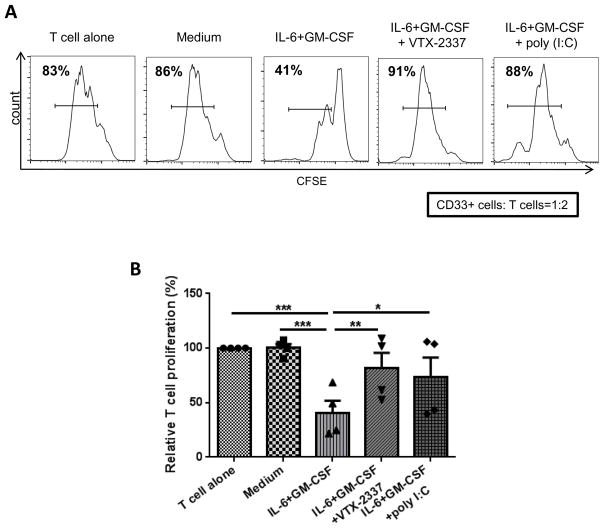
Figure 2.
HNSCC patient monocytes were cultured in T-75 flasks at 5x10⁵ cells/mL in complete medium with or without 10ng/mL GM-CSF and IL-6 for 7 days in the presence of motolimod or poly I:C. Medium and cytokines were refreshed every 2–3 d. Cells were then harvested and CD33+ cells purified by FACS sorting. Autologous T cells were isolated, labeled with CFSE and seeded in 96-well plates at 105/well. CD33+ cells from the above cultures were added to these cells at a 1:2 ratio. T cell stimulation was provided by anti-CD3/CD28 stimulation beads (bead:T cell = 1:1). Proliferation of T cells was analyzed by flow cytometry after 3 days. (A) Representative flow plot and (B) summary data are shown. Significance was calculated using one-way ANOVA followed by Tukey’s test, *p<0.05; **p<0.01.

