
Fig. 2. Human oral MSCs are highly susceptible for KSHV infection and viral latent infection leads to morphological and cell marker changes of MSCs.
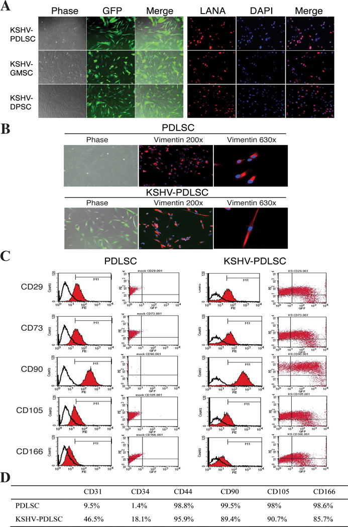
(A) Primary oral MSCs of different origins (PDLSCs, GMSCs and DPSCs) were infected with GFP-KSHV in an MOI of 50 (KSHV genome equivalent) for 48 hours and analyzed by GFP fluoresces. (B) Infected cells were drug-selected for a week followed by two weeks culture without selection and analyzed by IFA with anti-Vimentin antibody. Images show the phase-contrast, the antibody staining and GFP fluorescence of the cells. (C) Flow cytometric analysis of mock- and KSHV-infected PDLSCs with mesenchyme markers (CD29, CD73, CD90, VD105 and CD166, Y-axis) and GFP (KSHV-infected cells, X-axis). (D) The expression of mesenchymal and endothelial markers in mock- and KSHV-infected PDLSCs.