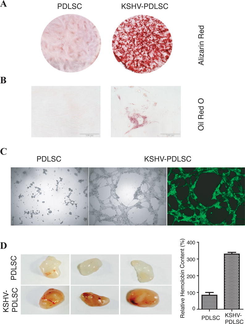
Fig. 3. KSHV infection promotes multi-lineage differentiation.

PDLSCs were mock- and KSHV-infected in an MOI of 50 (viral genomic DNA equivalent). (A) Cells were induced under osteogenic culture condition for 4 weeks and assayed for their osteogenic differentiation by Alizarin staining. (B) PDLSCs were subjected to adipogenic induction and adipogenic differentiation was analyzed by oil Red staining. (C) Mock- and virally infected PDLSCs were loaded on the top of Matrigel and the ability of the cells in formation of capillary-like tubules was analyzed under a ZEISS fluorescence microscope. (D) The effect of KSHV infection on angiogenesis property of PDLSCs was also examined ex vivo using the Matrigel plug assay. Matrigel containing mock- or KSHV-infected PDLSCs were subcutaneously implanted into C57BL/6 mice. After 7 days, Matrigel plug were removed and photographed. After Matrigel plugs were homogenized and centrifuged, their supernatant was used to quantitate the haemoglobin content using Drabkin’s reagent.
