
Fig. 4. Kidney capsule implantation of KSHV-infected MSCs.
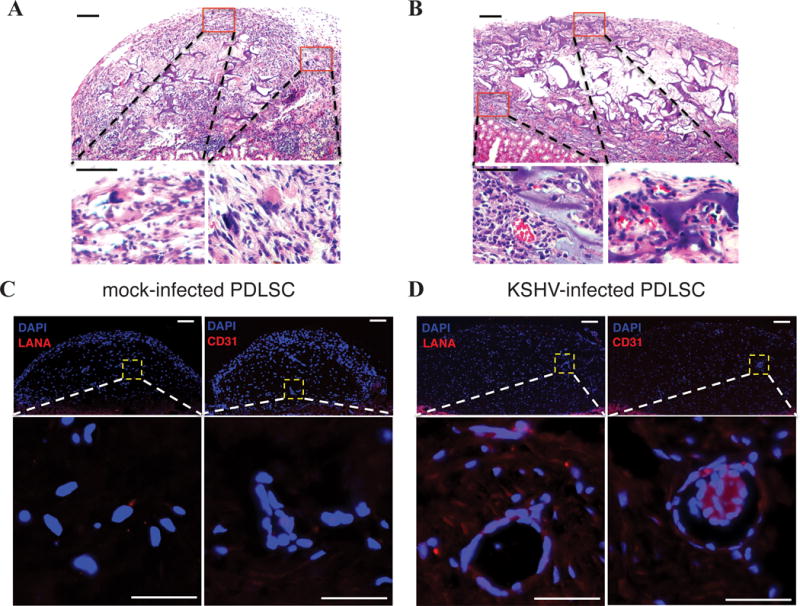
KSHV infected-PDLSCs (1×10⁶ cells) were implanted into kidney capsule. Spindle-like cells, sieve-like pattern, and mononuclear cells, with slit-like vascular spaces containing red blood cells were observed (B in comparison to A). Immunohistofluorescence staining showed expression of LANA and vascular endothelial marker CD31 in KSHV-infected human PDLSC transplant (B in comparison to A). Bar: 50 μm.