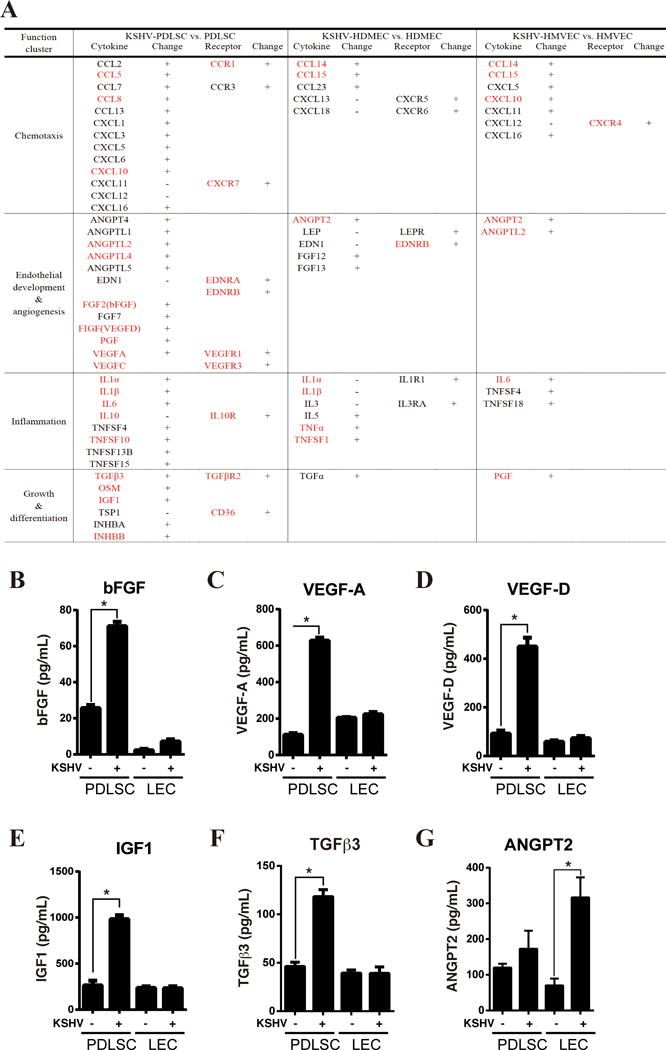
Fig. 5. Expression and secretion of chemokines and cytokines in PDLSCs and endothelial cells upon KSHV infection.

(A) Chemokines and cyckines that are up-regulated in KSHV-infected PDLSCs identified in our RNA-seq analysis are listed. Genes that were reported to be over-expressed in KS lesions are marked in red. The cytokines that are up-regulated in HDMEC and HMVECs upon KSHV infection are included as comparison. (B-G) PDLSCs and LECs were infected with KSHV in a MOI of 50 (viral genomic DNA equivalent). The infectivity rates of PDLSCs and LECs were determined using GFP fluorescence and were 92.1% and 88.5% respectively. Ninety-six hours post-infection, mock- and KSHV-infected cells were seeded in a relative low density (1×10⁵ per mL) with α-MEM containing 1% FBS. Supernatants were collected after 6 hours, and subjected to ELISA for bFGF, VEGF-A and VEGF-D, IGF1, TGFβ3 and ANGPT2.
