Figure 4.
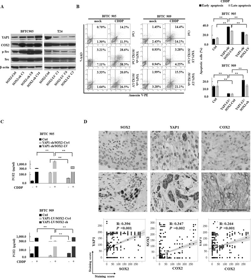
The regulation of YAP1 and COX2/PGE2 through the negative feedback of SOX2. (A) The expression levels of COX2, YAP1, and p-Src in SOX2-sh or SOX2-LV cells. (B) Apoptosis promoted by the inhibition of the YAP1-SOX2 axis. An apoptosis assay of BFTC 905 YAP1-sh/SOX2-LV cells and BFTC 909 YAP1-LV/SOX2-sh cells treated with CDDP for 72 h. Left, representative images of early apoptosis (bottom right quadrant) and late apoptosis (top right quadrant); Right, percentage of apoptotic cells. SOX2 induction recovered the anti-apoptotic ability attenuated by YAP1 knockdown, while SOX2 knockdown attenuated the anti-apoptotic ability protected by YAP1 induction. (C) An ELISA assay of PGE2 after treatment with CDDP for 72 h in YAP1-sh/SOX2-LV cells (upper) and YAP1-LV/SOX2-sh cells (lower). (D) The correlation among YAP1, COX2, and SOX2 in an immunohistochemistry analysis of 528 human primary UCB core tissues. Upper, representative images (scale bar, 500 µm); Lower, linear correlations among staining scores of YAP1, COX2, and SOX2.
Each error bar indicates mean ± SEM. **, P <0.01 (Kruskal–Wallis with post-hoc test [B and C]). See also Fig. S5 and S6.

