Figure 5.
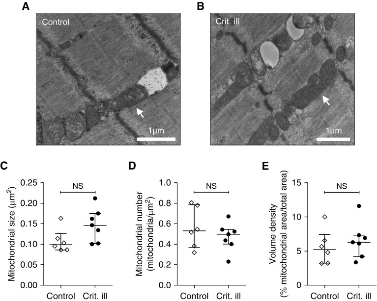
No major alterations of mitochondrial structure in diaphragm muscle fibers. (A) Electron micrograph of diaphragm biopsy from a control patient. Mitochondria are well aligned and have a normal general appearance. (B) Electron micrograph of a diaphragm biopsy from a critically ill patient. In A and B, the arrow indicates an example of a mitochondrion with a normal appearance. (C–E) Quantification of mitochondria in the micrographs revealed no differences in mitochondrial size, number, and density. Each data point represents the mean mitochondrial size/number/volume density as measured or counted in micrographs of a diaphragm biopsy of one patient. Data are presented as median and interquartile range. Crit. ill = critically ill; NS = not significant.

