Figure 1.

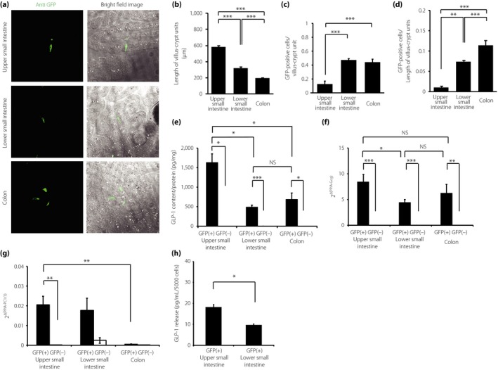
The density and quality of L cells in the gastrointestinal (GI) tract of glucagon (Gcg)‐green fluorescent protein (GFP) knock‐in (Gcg‐GFP) heterozygous (Gcggfp/+) mice. (a) Immunohistochemical images of the upper small intestine (UI), lower small intestine (LI) and colon of Gcggfp/+ mice (bright field image and fluorescence image). (b) The length of the crypt–villus units in the UI, LI and colon was measured by immunohistochemistry (n = 5). (c) The number of GFP‐positive (+) cells per crypt–villus unit (n = 5). (d) The density of GFP(+) cells was normalized against the length of the crypt–villus unit (n = 5). (e) Glucagon‐like peptide‐1 (GLP‐1) content in GFP(+) cells and GFP(–) cells from the UI, LI and colon (n = 5). (f, g) The messenger ribonucleic acid expression levels of Gcg and prohormone convertase (PC) 1/3 in GFP(+) and GFP(–) cells from the UI, LI, and colon (n = 5). Expression levels were normalized against the internal control, peptidylprolyl isomerase A (PPIA). (h) GLP‐1 secretion from GFP(+) cells from the UI and LI (n = 3). *P < 0.05, **P < 0.01, ***P < 0.001. NS, not significant.
