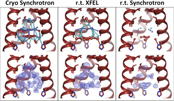
Fig. 1.
Low-pH (pH 5.5) structures of M2TM under cryogenic synchrotron (4QKC, Left), room temperature XFEL (5JOO, Center), and room temperature (r.t.) synchrotron (4QKM, Right) diffraction conditions. (Top) The front helix of each tetramer has been removed; waters are modeled as spheres, with red spheres representing full-occupancy waters and light and dark blue spheres as half-occupancy waters in alternate-occupancy networks A and B. Waters within hydrogen-bonding distance of each other are connected by sticks. The number of ordered waters decreases moving from Left to Right across the figure. (Bottom) Electron density for the pore solvent network (blue mesh) is shown to a contour of 0.5 σ. The largest amount of ordered density is present in the cryogenic synchrotron data collection condition. The volume and shape of the solvent density for the room temperature structures collected using XFEL and synchrotron sources are similar.

