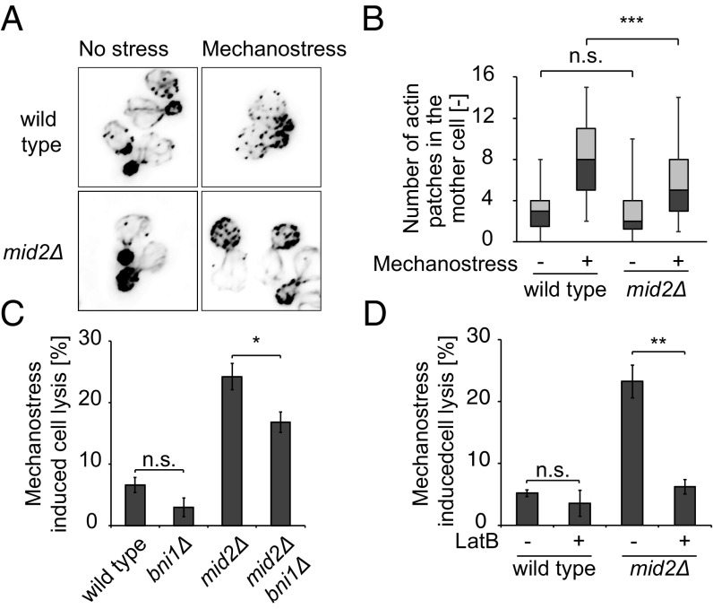
Fig. 4.
Inhibition of polarized growth prevents cell lysis upon mechanostress. (A) Wild-type or mid2Δ cells were synchronized and exposed (Right) or not (Left) to mechanostress for 30 min shortly after bud emergence. Actin cables and patches were then stained with rhodamine phalloidin. Images are taken with objective lens (magnification: 100×). (B) The number of actin patches in the mother cell was quantified in three independent experiments with at least 50 cells using a Fiji macro script from deconvolved images. The image quantification is represented as a box−whisker plot, where 5th, 25th, 50th, 75th, and 95th percentiles are depicted. Note that mechanostress signaled by Mid2 interferes with the assembly of a polarized actin network. (C) Deletion of the actin nucleator Bni1 partially rescues the cell lysis defect of mid2Δ cells. Exponentially growing wild-type and the indicated mutant cells were exposed to mechanostress for 3 h, and the percentage of cell lysis was quantified after trypan blue staining. The experiment was performed at least three times for each strain, with over 500 cells counted for each experiment. (D) Actin cables of exponentially growing wild-type and mid2Δ cells were depolymerized by treatment with 200 μM LatB for 15 min (+), before mechanostress was applied for 3 h. Control cells were treated with the solvent DMSO (−). The percentage of cell lysis was quantified after trypan blue staining from at least three experiments with more than 500 cells counted in each case. A Student’s t test was performed, and the significance is indicated [not significant (n.s.) = P > 0.05; *P ≤ 0.05; **P ≤ 0.01; ***P ≤ 0.001].

