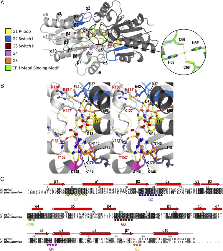
Fig. 1.
Crystal structure of KpUreG in complex with GMPPNP and nickel. (A) The structure of KpUreG (PDB ID code 5XKT) was solved as a dimer in complex with GMPPNP and nickel at a resolution of 1.8 Å. Cys66 and His68 from each of the two UreG protomers (colored in light and dark gray) coordinate a nickel ion in a square-planer geometry. Conserved motif (G1–G5) and CPH metal binding motif are colored as indicated. (B) A stereodiagram showing the interaction between KpUreG and GMPPNP. GMPPNP is sandwiched between the two KpUreG protomers and forms a network of hydrogen bonds (yellow dotted lines) with residues of the G1–G5 motifs. (C) Sequence alignment of KpUreG and HpUreG. The G1–G5 and the CPH metal-binding motifs are indicated as circles. Residues are numbered according to the HpUreG sequence. Apostrophes denote residues from the opposite protomer.

