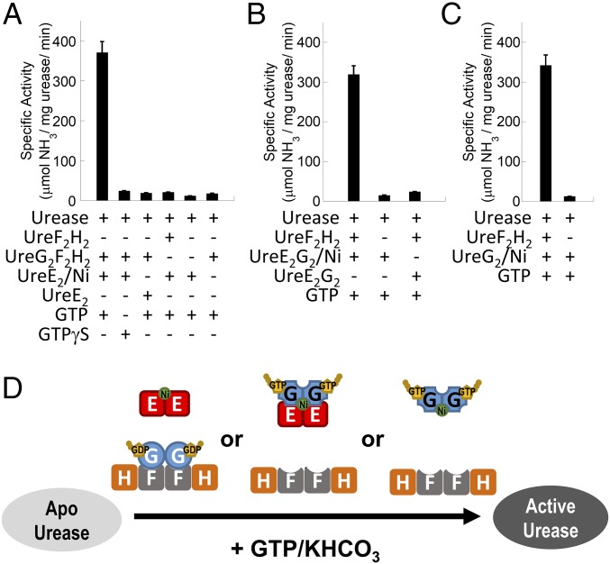
Fig. 5.
In vitro urease activation assays suggest that nickel-charged UreE dimer provides the nickel source for urease maturation. The in vitro urease activation assay was performed by incubating 10 µM H. pylori apourease with 20 µM of H. pylori urease accessory proteins/complexes as indicated at 37 °C for 20 min in 20 mM Hepes pH 7.5 buffer containing 1 mM GTP or GTPγS, 2 mM MgSO4, 10 mM potassium bicarbonate, 200 mM NaCl, and 1 mM TCEP. Urease activity was measured by the amount of ammonia released. Protein samples of urease accessory proteins/complexes were prepared and analyzed by SEC/SLS (SI Appendix, Fig. S8). Nickel-charged UreE dimer (UreE2/Ni) and nickel-charged UreE2G2 (UreE2G2/Ni) complex were prepared and analyzed by atomic absorption spectroscopy (SI Appendix, Fig. S5). (A) Apourease was activated only when 20 µM nickel-charged UreE dimer, providing the sole source of nickel, was added with the presence of 20 µM UreG2F2H2 complex. (B) Apourease was activated when 20 µM nickel-charged UreE2G2 complex (UreE2G2/Ni), providing the sole source of nickel, was added with the presence of 20 µM UreF2H2 complex. (C) Apourease (10 µM) was activated when 20 µM nickel-charged UreG dimer (UreG2/Ni), providing the sole source of nickel, was added with the presence of 20 µM UreF2H2 complex. (D) Schematic diagram summarizing the combination of urease accessory proteins/complexes that can activate urease in the in vitro assay. Either UreE2/Ni, UreE2G2/Ni, or UreG2/Ni can provide the nickel source for urease activation.

