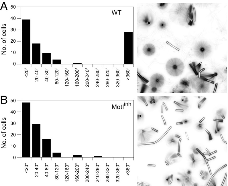
Fig. 2.
MotI abolishes powered flagellar rotation. Wild-type strain DS8022 cells (A) and MotIInh strain DK1995 cells (B) were tethered by flagellar stubs and monitored for 60 s in time-lapse microscopy. (Left) The angles of rotation of 100 cells were binned and expressed as a frequency histogram. (Right) Time-lapse composite imaged of sample fields. Sample movies used to generate these data are included as Movie S1 (wild-type) and Movie S2 (MotIInh) in Supporting Information.

