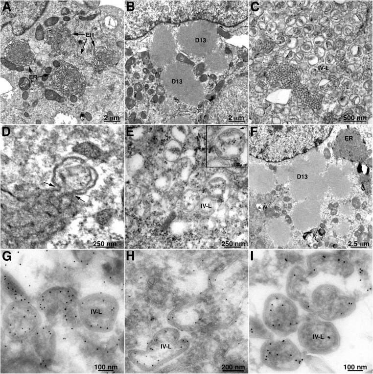
Fig. 5.
Modification of ER membranes by expression of VACV A17 and D13 proteins. BS-C-1 cells were infected with a recombinant VACV expressing T7 RNA polymerase (vTF7-3) in the presence of the DNA replication inhibitor AraC and transfected with plasmids with T7 promoters regulating (A) full-length A17 alone, showing tightly curved ER networks; (B) D13 alone, showing protein inclusions; (C and D) D13 and full-length A17, showing IV-L structures with arrows pointing to membrane junctions; (E) D13 and C-terminal truncated A17, showing IV-L structures; (F) D13 and N-terminal truncated A17, showing D13 inclusions and tightly curved ER networks in the same cell. The bottom row contains images of BS-C-1 cells infected with vTF7-3 in the presence of AraC and transfected with plasmids expressing D13 and full-length A17. Cells were cryosectioned and stained with antibody to (G) A17, (H) D13, and (I) protein disulfide isomerase and then protein A conjugated to 10 nm gold.

