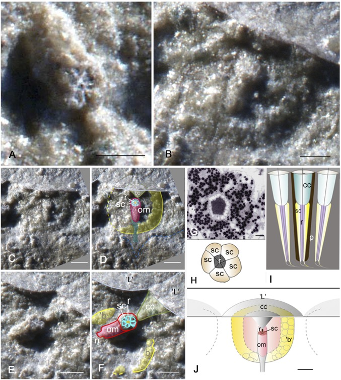
Fig. 2.
Internal structures of the functional visual unit. (A) Ommatidium. Note the cellular elements (relicts of receptor cells) arranged radially around the central core (relict of the rhabdom). (B) Ommatidium positioned in a basket. Note the cellular elements (relicts of receptor cells) arranged radially around the central core (relict of the rhabdom). (C) General aspect of B for interpretation in D. (E) General aspect of A for interpretation in F. (G) Cross-section of the ommatidium of the extant crustacean Dulichia porrecta (Bate, 1857) (87) (Crustacea, Amphipoda) (88). (H) Schematic drawing of the elements of a typical sensory system in the aquatic compound eye in G. (I) Schematic drawing of a longitudinal section of an ommatidium. (J) Schematic drawing of the visual unit of S. reetae. b, basket; cc, crystalline cone; L, lens; om, ommatidium; p, pigment screen; r, rhabdom; sc, sensory (receptor) cells. (Scale bars: A, B, E, F, and J, 200 μm; C and D, 100 μm; and G, 1 μm.)

