
Figure 1.
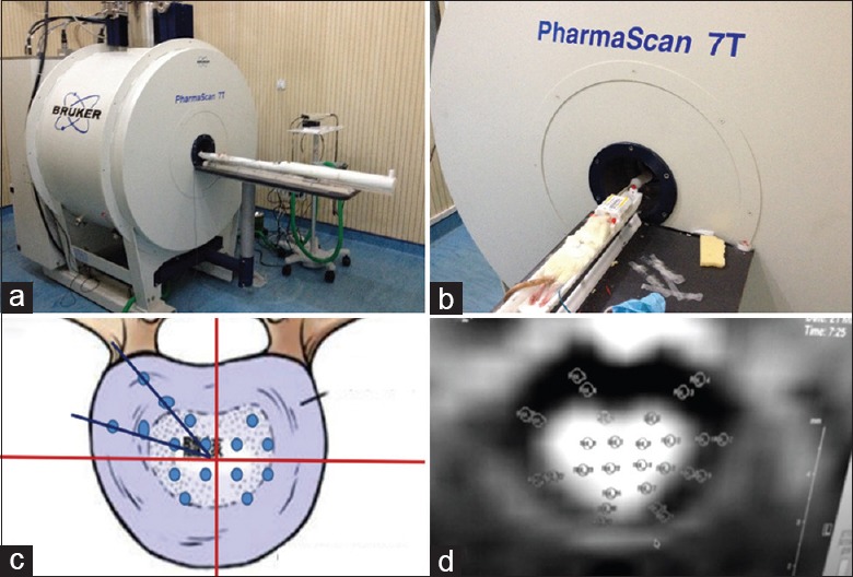
(a) 7.0T Magnetic resonance imaging and (b) body position in the magnetic resonance imaging scanning. Twenty-eight images were obtained of each imaged layer. (c) The disc (including nucleus pulposus and annulus fibrosis) was divided into four quadrants of equal area. (d) Due to uncertainty in differences regarding the distribution of fractional anisotropy values, regions of interests were manually determined from the outer annulus fibrosis, inner annulus fibrosis, and nucleus pulposus areas of each quadrant for the purpose of statistical analysis and image reconstruction.